Transduction
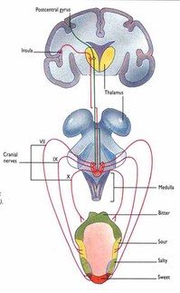
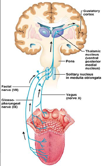
The chemistry of taste does not simply stop at the taste bud! After different molecules either enter through their specific ion channels or latch on to their specific taste receptor complex, the action potential created in the taste cells must then be relayed to the brain where it is interpreted. Directly in each and every taste bud is the terminal of a primary taste sensory neuron. Through electrochemical signals, the stimulation is carried from these sensory neurons, all the way to two major regions in the brain known as the limbic system and the cerebral cortex. Depending on the region in which each taste bud is located, there are primarily three different nerves that the taste cells synapse with to carry the message to the brain. First of all, the chorda tympani (VI) is the nerve that conducts signals from taste buds located in the front of the tongue and on the sides of the tongue. These are more commonly found to be sour, sweet and salty tastes, but are not limited to bitter and umami tasting molecules too. Furthermore, taste buds on the back of the tongue, which are most commonly receptive to bitter tasting molecules, are conducted to the brain by the glosso-pharyngeal nerve (IX). Last but not least, the vangus nerve (X) is responsible for transmitting signals from the roof of the mouth and the larynx. Each of these three cranial nerves later synapse in the brainstem in the “nucleus solitary tract” before continuing to the thalamus and different regions of the brain.
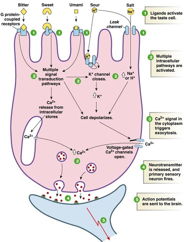
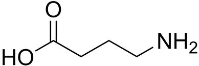
In order for the afferent nerves to be activated, a chemical signal bust be passed from the taste cells to either the chorda tympani, glossopharyngeal or vangus nerve. This takes place over a synapse, which is an extremely small gap between the two nerve cells. In order for the signal to be conducted, a neurotransmitter must diffuse from the taste cell to the afferent nerve. (see youtube video below for synapse animation) For this to happen, the taste cells in which an action potential is created in, contains groups of neurotransmitters held together in synaptic vesicles. In the case of the gustatory system, the excitatory neurotransmitters happen to be the amino acid, glutamate. Thus it is glutamate that causes the depolarization of the postsynaptic membrane. (see steps 4 and 5 in the figure below to the left) The synaptic vesicles cluster on the inside of the cell membrane. This is considered to be the presynaptic side of the synapse. When an action potential is created in the taste cell (from the stimulation as a result of the binding of a sweet, sour, bitter, salty or umami molecule to its receptor or through its ion channel), the neurotransmitters are released to diffuse across the synaptic cleft, which is the space found in between the taste cell and the afferent neuron. From there, the neurotransmitters (glutamate) bind on to receptors found on the postsynaptic membrane to trigger a new action potential in the afferent nerves. By binding to these receptors, the new action potential is similarly created as positive ion channels are signaled to be opened and the cell becomes depolarized. Just as the food molecules binded to the taste receptors and created a depolarization of a channel, this depolarization then moves along the nerve in the exact same manner as the positive charges both inside and outside the membrane are attracted to each other. However, the inhibitory amino acid neurotransmitter, known as gamma amino butyric acid (see figure below to the right), works to slow down or stop the action potential by opening negative ion channels to hyperpolarize the post synaptic membrane and inhibit the action potential. Alike many similar molecules, this acid is water soluble as a result of its polarity from the carboxyl and amino group on each end. Hydrogen bonds can readily be made with water, and a lot of energy must be added to the molecule to break its bonds. Therefore, from this point, the action potential travels along the three afferent nerves from the tongue to the brainstem.
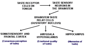
From the brainstem, in which the nucleus of the solitary tract (gustatory nucleus) is located, signals are then relayed by axons to the thalamus where they are further projected to the to the gustatory cortex which is located in the cerebral cortex. The gustatory complex, which is surrounded by the Insula and the frontal operculum, is responsible for perceiving the tastes. Additionally, information from the olfactory cortex and somatosensory cortex, which happen to be the smell and texture systems, then combine with the perception of taste to create and interpretation of the food. Furthermore, a combination of all of this information is sent via the chemical depolarization of nerve cells as explained earlier, to the limbic system and the orbitofrontal cortex. The limbic system includes parts of the brain such as the hippocampus, the hypothalamus and the amygdala. It is in these areas that memory begins to form in regards to the things that we eat, as well as emotions suited to the memory. Specifically, the amaygdala and hypothalamus are in charge of the “emotional quality of taste” and the hippocampus is in charge of memory. The gustatory area of the cerebral cortex on the other hand is what perceives the tastes. diagram pic As a result, a feedback loop formed as foods either positively or negatively stimulate these different areas of the brain. Thus, using electrochemical signals to further relay a message to the individual to stop eating the food, or to keep eating the food. Since much of the limbic system is associated with memory, each food that the person has already experienced, will be familiar to the gustatory system.